Fig. 3.
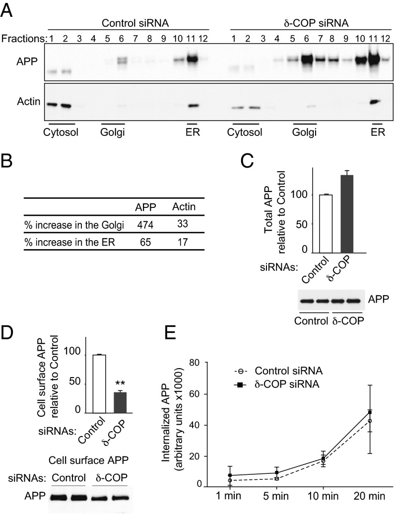
Effect of δ-COP on APP localization. (A) Subcellular localization of APP and actin in N2a-695 cells transfected with control siRNA (Left) or δ-COP siRNA (Right) by sucrose density gradient fractionation. (B) Protein expression was evaluated by Western blotting analysis and quantified 48 h posttransfection with δ-COP siRNA. (C) Total APP measurement following transfection with δ-COP siRNA by Western blotting analysis (Lower) and quantification (Upper). (D) Cell surface expression of APP was analyzed by Western blotting analysis (Lower) and quantified using cell surface biotinylation (Upper). (E) Biotinylation experiments were conducted to study APP internalization in N2a-695 cells transfected with δ-COP siRNA. Biotinylated proteins were internalized at 37 °C for different amounts of time (1 min, 5 min, 10 min, and 20 min). The internalized APP was analyzed by Western blotting and quantified (**P < 0.01, two-tailed Student’s t test; n = 3).

