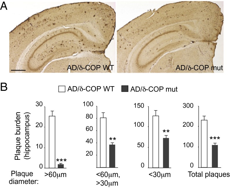
Fig. 3.
Effect of partial inactivation of δ-COP on amyloid plaque formation evaluated by immunohistochemistry in the hippocampus of 2xTg AD mice. (A) Representative images show amyloid plaques in the hippocampus of male mice at 9 mo (n = 6 per genotype). (Scale bar, 500 μm.) (B) Quantification of the number of plaques in the hippocampus (n = 6 per genotype). (**P < 0.01, ***P < 0.001, two- or one-tailed Student’s t test with or without Welch’s correction. The results are shown as means ± SEM.)

