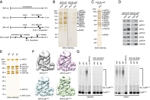
Fig. 1.
Immunopurification of mitotically phosphorylated APC/CCDC20 and purification and characterization of nonphosphorylatable and phospho-mimicking human APC/C. (A) Schematic overview of the synchronization procedures to obtain mitotic HeLa S3 cells. To obtain prometaphase-arrested cells with active SAC (SAC on), cells were synchronized using either the spindle poisons nocodazole or taxol. To obtain metaphase-arrested cells with inactive SAC (SAC off), cells were synchronized using taxol and supplemented with the Aurora B inhibitor hesperadin. Cells exposed to taxol with or without hesperidin were also treated with the proteasome inhibitor MG132 to prevent mitotic exit (41). To enrich for mitotic cells with unperturbed spindle checkpoint, cells were synchronized using a double-thymidine arrest–release procedure (SAC on/off). Mitotic cells were harvested by mitotic shake-off. (B–D) APC/C was immunoprecipitated from extracts of asynchronous (Asyn) or nocodazole-arrested (Noc) HeLa cells using APC3 antibody beads, re-immunoprecipitated using CDC20 antibody beads, and subjected to SDS/PAGE and silver staining (B and C) or Western blotting (D). Positions of subunits that display a mitotic phosphorylation electrophoretic mobility shift are indicated (pAPC1, pAPC3, and pAPC8). Note that in the CDC20 re-IP only the slowly migrating forms of APC1, APC3, and ACP8 were detected and that these three all cross-reacted with phospho-specific antibodies to these subunits. (E) Silver-stained SDS/PAGE gel of purified recombinant WT, nonphosphorylatable (pA), and phospho-mimicking (pE) APC/C. (F) Single-particle reconstruction by negative stain EM of recombinant APC/Ccoactivator–UBCH10–Ub–substrate complexes: APC/CCDH1 (gray, 20 Å resolution) (7), APC/C-pACDH1 (pink, 16 Å resolution), APC/C-pECDH1 (blue, 16 Å resolution), and APC/C-pECDC20 (green, 19 Å resolution). pA and pE maintain APC/C structural integrity. (G) Ubiquitination reactions, carried out in the presence of recombinant APC/C-WT, pA or pE, and fluorescein-labeled substrate N-terminal fragment of cyclin B (CycBNTD*), were analyzed by SDS/PAGE and fluorescence scanning. The reactions with CDC20 with or without MCC are in the Left panel, and the ones with CDH1 with or without EMI1 (EMI1-SKP1) are in the Right panel.

