
Figure 3.
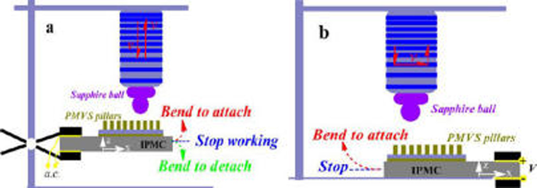
Schematic illustration for detecting normal (a) and shear (b) adhesions. A rigid sapphire ball was fixed at the end of force sensor to act as the upper mating ball, and the IPMC membrane carrying an array of PMVS micropillars was used as the sample. The IPMC was auctated by a low voltage of 1.0, 1.5, or 2.0 V.