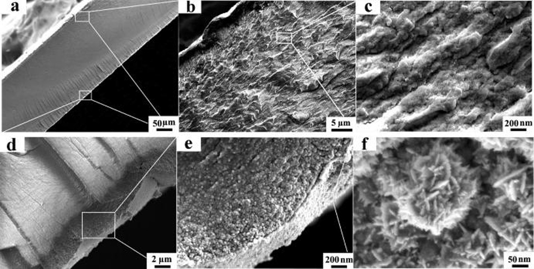
Figure 6.

SEM images of the IPMC. After freeze cracking in liquid N2, samples were deposited with Pt particles (~5 nm) for SEM imaging. (a) Cross-sectional image of the IPMC at a magnification of 200×. The two surfaces are sandwiched between two layers of Pt nanosheets. (b, c) Cross-sectional image of the hybrid membrane with magnifications of 3000× (b) and 50 000× (c). (d, e) Pt nanosheet with magnifications of 5000× (d) and 50 000× (e). (f) Top-view image of Pt nanograins with a magnification of 200 000×.
