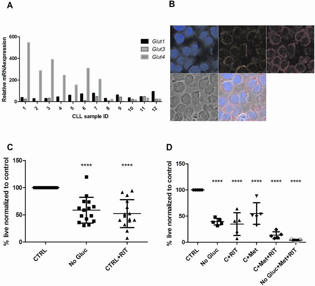
Fig 3. CLL cells express elevated GLUT4 that exhibits plasma membrane co-localization and treatment with GLUT4 targeting agent ritonavir, elicits apoptosis.
A – Expression of GLUT1, GLUT3, GLUT4 in patient samples and normal B lymphocytes by qRT-PCR. Relative quantification is normalized to expression in NBL (not shown). B – GLUT4 plasma membrane localization in CLL cells; clockwise from top left- DAPI, wheat germ agglutinin stain of plasma membrane, GLUT4, phase contrast and Merge. C - CLL patient cells (n=15) cultured glucose-containing media or glucose-free media, in the presence or absence of 20µm ritonavir. Cell viability is determined by AnnexinV and DAPI staining. All samples were normalized to viability of CLL patient cells cultured in 5mM glucose –containing media. D - CLL patient cells (n=5) cultured in glucose-containing and glucose-free media and treated with 20µm ritonavir, 5mM metformin and the combination for 48hours. Cell viability measurement and normalization as in C. ****p <0.0001.

