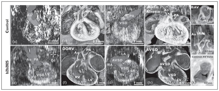
FIGURE 1.
Ultrasound diagnoses of CHD and cilia defects in CHD mutants. (a and b) Vevo 2100 color flow imaging showed crisscrossing of blood flow indicating normal aorta (Ao) and pulmonary artery alignment, confirmed by histopathology (b). Cd, caudal; Cr, cranial; L, left; LV, left ventricle; R, right; RV, right ventricle. (c and d) Embryonic day (e) 16.5 mutant mouse (line b2b327) exhibited a blood flow pattern indicating single great artery (pulmonary artery) and ventricular septal defect (VSD) (c), suggesting aortic atresia with ventricular septal defect, confirmed by histopathology (d). (e–h) Color flow imaging of E15.5 mutant mouse (line b2b2025) with heterotaxy (stomach on right) showed side by side aorta and pulmonary artery, with the aorta emerging from the right ventricle, indicating DORV/ventricular septal defect (e and f) and the presence of AVSD (g and h). AVSD, atrioventricular septal defect; DORV, double outlet right ventricle; LA, left atrium; RA, right atrium. (i–k) Histopathology also showed a bicuspid aortic valve (BAV) (i), interrupted aortic arch (IAA) (j), and common atrioventricular valve (k). Reproduced with permission from [9▪▪].

