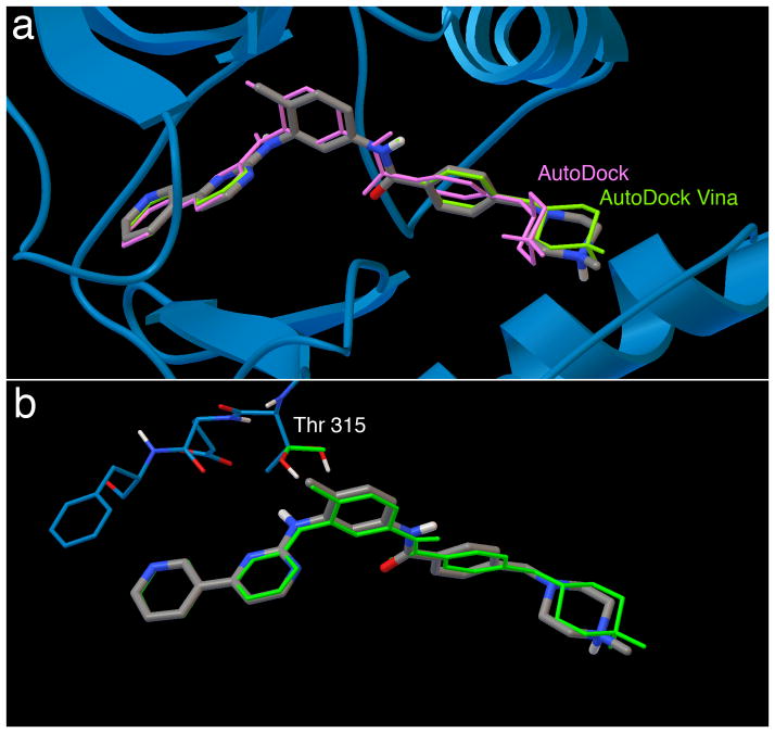
Figure 2. Results of docking imatinib to its receptor in bound and apo conformations.
a. Redocking of flexible imatinib to rigid Abl (PDB entry 1iep) using AutoDock (purple) and AutoDock Vina (green). The X-ray crystallographic ligand position is in silver.
b. Cross docking of flexible imatinib to Abl (PDB entry 1fpu) with a single flexible residue side chain using AutoDock Vina (green). Note that Vina does not retain hydrogen atom positions during docking, so the threonine hydroxyl hydrogen is placed in a random position in the docked coordinate set.

