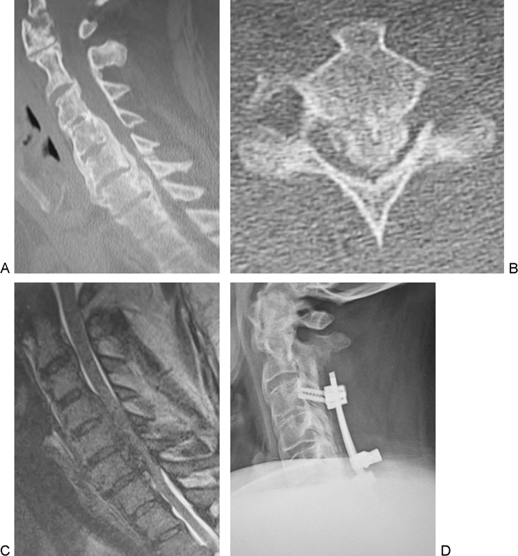
Fig. 2.

Case 6. Sagittal (A) and axial (B) computed tomography shows diffuse idiopathic spinal hyperostosis (C2–C5, C7–T2) and mixed-type ossification of the posterior longitudinal ligament(C1–C2, C4–C7). Sagittal image on T2-weighted magnetic resonance imaging (C) shows severe cord compression. Postoperative plain lateral radiograph (D) shows postoperative posterior spinal instrumentation and fusion (C3–C7) with laminectomy (C2–C7).
