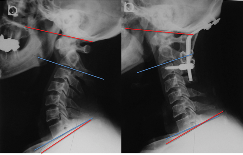
Fig. 4.

Case 1. A 54-year-old man with os odontoideum. Preoperative (left) and 3 years postoperative (right) X-rays. Occipitocervical kyphosis was corrected and lordosis gained at 3 years postoperatively. Conversely, subaxial lordosis decreased, and the McGregor and T1 slopes were maintained.
