Figure 1.

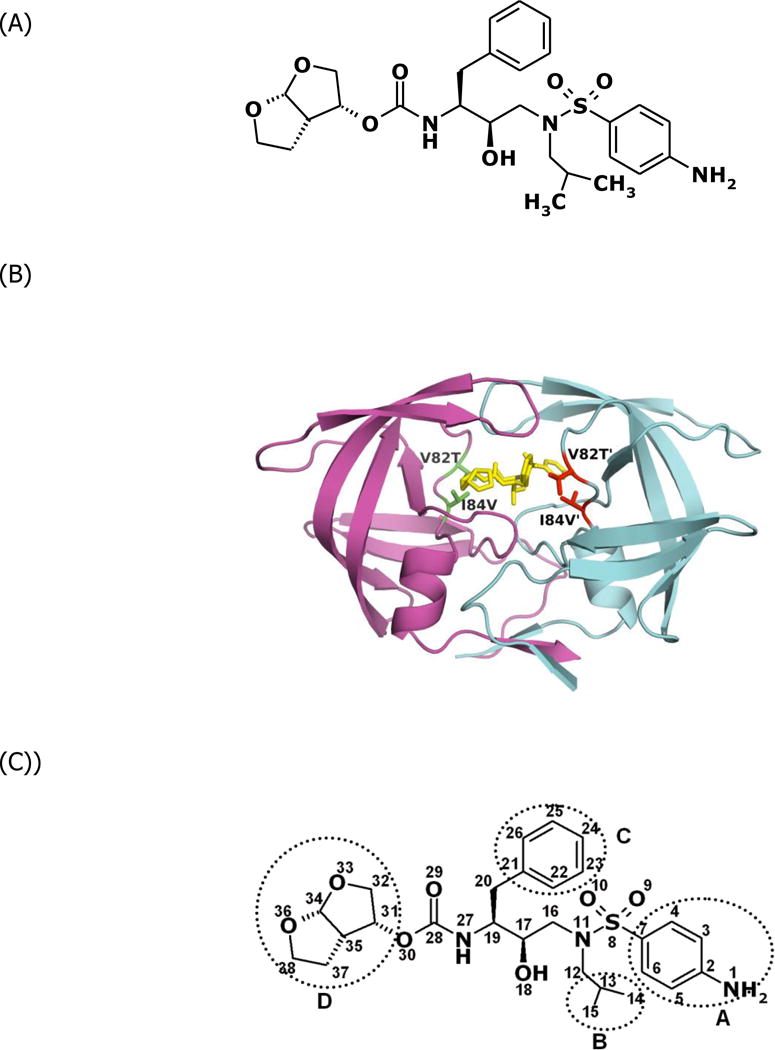
(A) Chemical structure of DRV. (B) Structure of protease variant ACT-DRV complex. DRV is colored yellow. The side chains of the mutated residues Thr82 and Val84 are displayed and colored red or green. (C) The four moieties of DRV.

(A) Chemical structure of DRV. (B) Structure of protease variant ACT-DRV complex. DRV is colored yellow. The side chains of the mutated residues Thr82 and Val84 are displayed and colored red or green. (C) The four moieties of DRV.