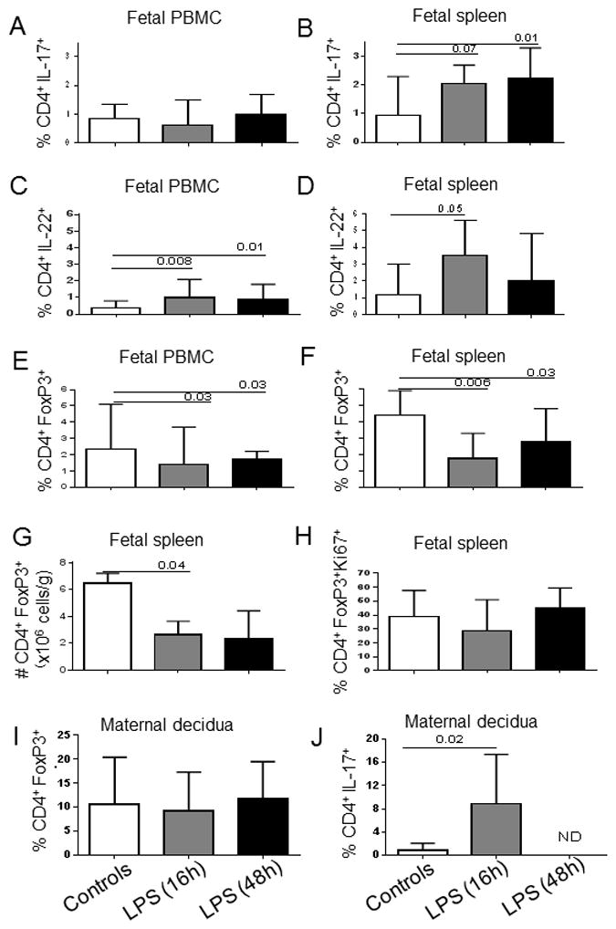
Figure 1. Intra-amniotic LPS alter Th17 (IL-17 and IL-22) and Treg response by fetal CD4 T-cells.

Bars show the median (range) percentage of CD3+CD4+ T-cells from PBMC and spleen expressing IL-17 (A, B), IL-22 (C, D) or FoxP3 (E, F). G) Absolute counts of splenic Treg (CD3+CD4+FoxP3+). H) Percentage of splenic Treg expressing Ki67 within the CD4+FoxP3+ population. Control group (n=8) and LPS-exposed fetuses for 16h (n=6) and 48h (n=8). I) Percentage of decidua Treg (CD3+CD4+FoxP3+) within the CD4+ T cell population. Decidua from unexposed dams (n=8) and from those exposed to IA LPS for 16h (n=4) and 48h (n=7). J) Percentages of decidua CD3+CD4+ T-cells expressing IL-17. Control group (n=4) and decidua from dams exposed to IA LPS for 16h (n=4). N.D.: Not done. Significant differences between groups were calculated using Mann-Whitney U tests test. Bars graphs show the median and interquartile range.
