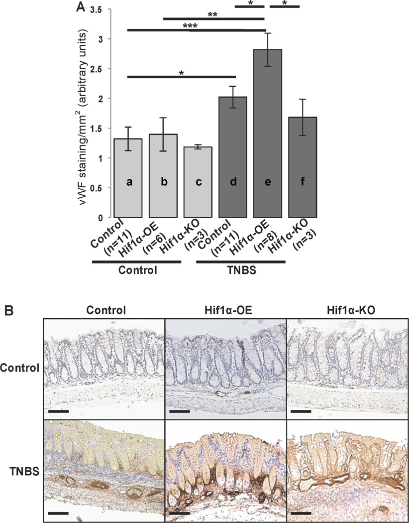
Figure 5.
HIF-1α-OE, HIF-1α-KO and control littermates were treated as indicated with TNBS (250mg/kg). Mice were sacrificed 48 h later and colon tissues were fixed in formalin, embedded in paraffin and endothelial cells where stained with vWF antibody (n=3 to 11). (A) Relative colon tissue angiogenesis (vWF positive cells/mm2) quantified by AxioVision (Carl Zeiss microscopy) in HIF-1α-OE, HIF-1α-KO and control littermates treated as indicated with TNBS (250mg/kg). Statistical analysis was performed using student’s t test. *p<0.05 (B) Representative images from vWF stained colon tissues sections. Scale: 100 µm.

