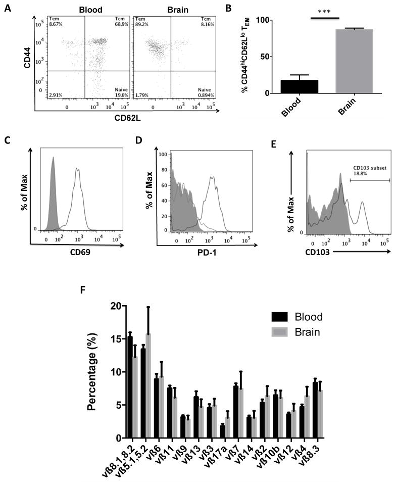
Figure 4. CNS-resident CD8 T cells stochastically migrate into the brain and have tissue-resident and effector memory phenotypes.
A representative dot plot shows effector memory phenotype of CD8 T cells in the aged brain and central memory phenotype of CD8 T cells in the aged blood (A). The percentage of CD8 T cells with effector memory phenotype (CD44hiCD62lo) was quantified (B). Representative histograms (N= 5–6/group) depict significant expression of the activation markers CD69 (C) and PD-1 (D) in the brain (solid black) relative to blood (solid gray), as well as the resident memory marker CD103 (E). A mouse vβ TCR screening panel containing 15 FITC-conjugated monoclonal antibodies was used to assess TCR vβ usage in CD8 T cells from the blood and brain of aged mice. The mean percentage for each of the 15 subfamilies of T cell receptor vβ is displayed (F). No differences were found between groups (N= 11/group). Cell-specific FMO controls were used to determine positive gating (shaded gray). Error bars show mean SEM. Abbreviation: SEM standard error of mean. *p<0.05; **p<0.01; ***p<0.001

