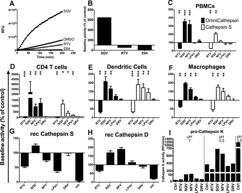
FIGURE 1. HIV PIs variably alter cathepsin activities in human CD4 T cells, DCs and macrophages.
(A) Omnicathepsin activity was monitored with fluorogenic substrate every 5 min in live PBMCs pretreated with DMSO, 5μM RTV, 5μM SQV or 10μM E64. (B) The maximum slope of fluorescence emission over 1 h in the presence of DMSO was represented as 100% and the effect of each PI was calculated as % of control. (C) PBMCs, (D) CD4 T cells, (E) DCs or (F) macrophages were pretreated for 30min with DMSO (control) or with 5μM of indicated PIs before adding specific cathepsin substrate (Omnicathepsin black bars, Cathepsin S white bars for C, D, E and F). Data represent average +/− SD of cells from 6 healthy donors. (G) Recombinant cathepsin S or (H) recombinant cathepsin D were pretreated with DMSO or 5μM of indicated PIs or inhibitor (10μM ZFL-COCHOO for cathepsin S, 100μM Pepstatin A for cathepsin D) before adding specific substrate for each activity. In each panel, 100% represents the maximum slope of DMSO-treated enzyme. Data represent average +/− SD of 3 independent experiments. (I) Pro-cathepsin K was incubated with 5 μM of DMSO or indicated PIs for 30min at different pH before adding specific substrate. pH 3.5 was used to trigger the maximal procathepsin maturation. The maximum slope of fluorescence emission is represented. *p< 0.05, **p< 0.01, ***p<0.001.

