
Figure 3. Analysis of Mcl-1's putative D-box.
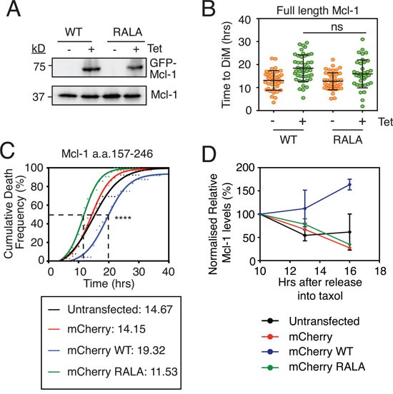
A. Immunoblot of endogenous Mcl-1 (lower panel) and GFP-tagged Mcl1 (upper panel) following 24-hour 1 μg/ml tetracycline induction. B. Quantitation of time to mitotic death of RKO cells expressing Mcl-1WT and Mcl-1RALA following incubation with 0.1μM taxol and 1 μg/ml tetracycline. Zero hours represents mitotic entry. Mann Whitney U test, ns p > 0.05. C. Cumulative death frequency of RKO Cyclin B1 R42A cells transiently transfected with pLNCX2 plasmids expressing myc-tagged mCherry fused to Mcl-1 competitor fragments (a.a. 157-246) with or without the RALA mutation. Transfected cells were identified by fluorescent microscopy and tracked by phase contrast microscopy. Mann Whitney U test between mCherry WT and mCherry RALA, **** p < 0.0001. D. Quantitation of two independent immunoblots showing relative Mcl-1 levels in RKO Cyclin B1 R42A cells transiently transfected with the mCherry-Mcl-1 WT and RALA fragments exposed to 0.1 μM taxol for the times indicated.