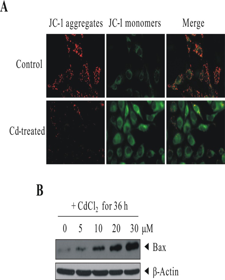
Figure 4. Cd treatment induced the loss of mitochondrial transmembrane potential and the up-regulation of proapoptotic protein BAX.
(A) BEAS-2B cells were sham-exposed or dosed with 30 μM CdCl2 for 18 h. JC-1 assay was conducted as described in “Materials and methods” for the determination of mitochondrial membrane integrity. (B) BEAS-2B cells were sham-exposed or dosed with increasing concentrations of CdCl2 for 36 h; cells were lysed; and protein extracts were subjected to western blot analysis using antibodies against BAX. The same blot was stripped and reprobed with the monoclonal β-actin antibody to monitor the loading difference. The results are representative of three independent experiments.

