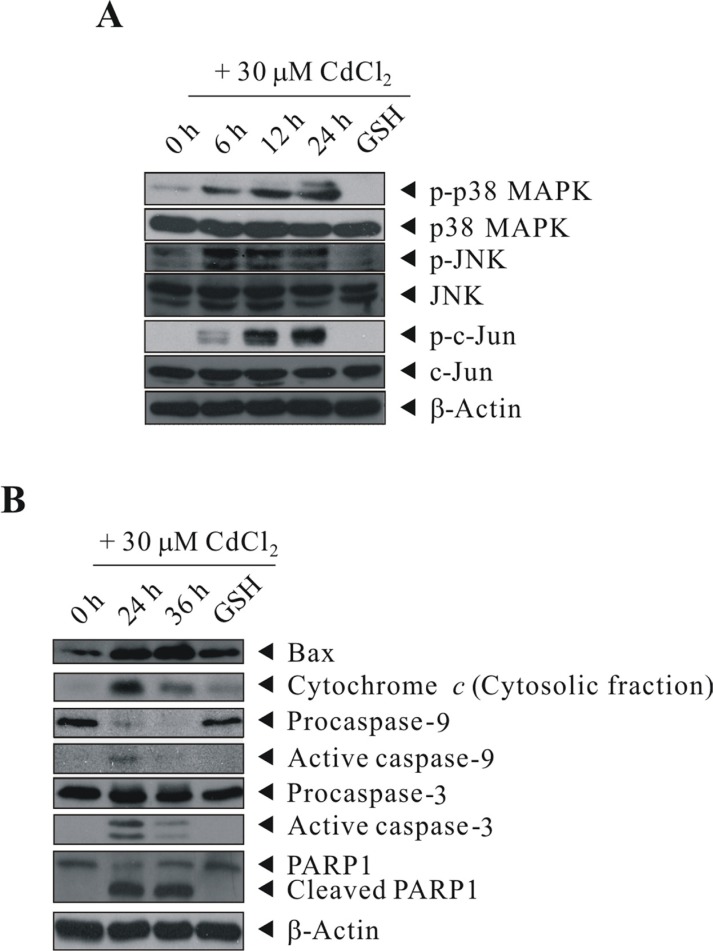
Figure 5. GSH inhibited the activation of Cd-induced p38 MAPK/JNK pathways and proteins involved in apoptosis signaling.
BEAS-2B cells were sham-exposed or dosed with 30 μM CdCl2 for 6, 12, or 24 h (A) or with 30 μM CdCl2 for 24 or 36 h (B); cells were lysed; and protein extracts were subjected to western blot analyses using antibodies against p-p38 MAPK, p38 MAPK, p-JNK, JNK, p-c-Jun, c-Jun, Bax, Cytochrome c, Caspase-9, Caspase-3, and PARP1. Cells were also pretreated with 20 mM GSH for 1 h before exposed to 30 μM CdCl2 for 24 (A) or 36 h (B). The same blot was stripped and reprobed with the monoclonal β-actin antibody to monitor the loading difference. The results are representative of three independent experiments.

