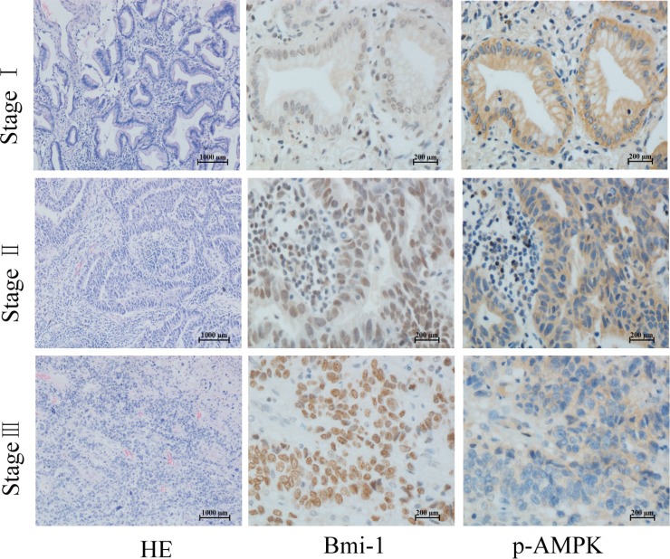
Figure 2. Phosphorylation of AMPK and Bmi-1 expression in lung cancer.
Paraffin tissue blocks were sectioned from 65 lung adenocarcinoma specimens at different pathological stages and stained with antibodies as described for Figure 1, and counterstained with hematoxylin and eosin (HE). Representative images are shown.

