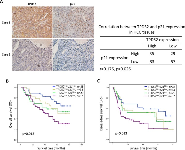
Figure 5. Immunohistochemical analysis for the correlation between TPD52 and p21 protein expression.
A. Immunohistochemical staining of TPD52 or p21 was performed in the serial sections from the same tumor tissues. A summary of the results was shown that there is significant positive correlation between TPD52 and p21 expression in HCC tissues (p = 0.026). B. and C. High expression of both TPD52 and p21 indicated better overall survival and disease-free survival of patients with HCC. (A, ×200 magnification).

