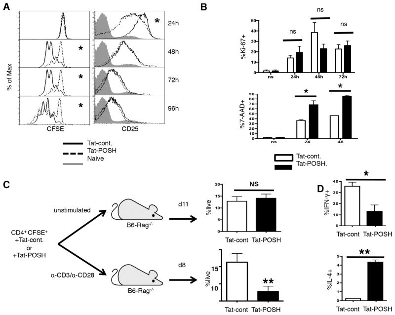
Figure 1. POSH regulates CD4+ T helper differentiation and survival.
(A) Naïve CD4+ T cells were labeled with CFSE, treated with Tat-POSH or Tat-cont. and stimulated with α-CD3 and α-CD28 in the presence of IL-2 and Tat-POSH or cont. for 4 days. Cell division was analyzed by dilution of the CFSE dye (left) and the level of CD25 (right) was determined by flow cytometry. (B) Naïve CD4+ T cells were treated with Tat-POSH or Tat-cont. and stimulated for 24, 48 and 72 hours with exogenous IL-2 and the percent of Ki-67+ cells (top) and 7-AAD+ (bottom) cells was determined. All experiments are representative of n>4 independent experiments. (C) Naïve CD4+ T cells were labeled with CFSE and treated with Tat-POSH or Tat-cont. for 30 minutes. 1×10⁶ cells were either un-stimulated or stimulated with α-CD3 and α-CD28 then adoptively transferred into Rag−/− mice. Cell numbers and division status were assessed in the spleen and lymph nodes at day 11 and day 8. n=3 independent experiments with cohorts of 5 mice per condition per experiment. (D) Naïve CD4+ T cells were treated as in (A) and re-stimulated for 5 hours with PMA/Iono in the presence of BFA n=4. The percent of IL-4+ and IFN-γ+ cells were then determined by ICCS. All graphs show +/− SD * indicates p < 0.05; ** p < 0.01.

