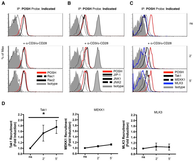
Figure 5. Composition of the POSH scaffold complex in unique in CD4+ T cells.
(A–D) Naïve CD4+ T cells were stimulated with cross-linked α-CD3 and α-CD28 for 2 or 5 min. Lysates were subjected to IP-FCM using α-POSH CML beads and probed with Rac1 and Rac2 (A), JIP-1, JNK1 and JNK2 (B), or Tak1, MEKK1, and MLK3 (C). Probing with α-POSH was used to confirm pull-down and serve as a ‘loading control’ and an isotype is used as a negative control. (D) The fold induction of MAP3K-POSH interactions over non-stimulated (ns) was calculated. All experiments are representative of >3 independent experiments. * indicates p < 0.05.

