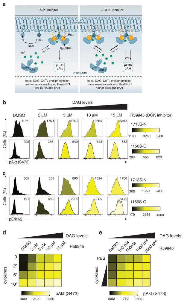
Figure 4.
RasGRP1 couples to the cytokine receptor via basal DAG. (a) Model of RasGRP1 in basal state. (Left) RasGRP1 exists in autoinhibited form but can use basal Ca2+, DAG to shuttle between the cytoplasm and membrane. DGK converts DAG to PA (phosphatidic acid). (Right) In the presence of DGK inhibitor, DAG is no longer converted to PA and accumulates at the membrane. As a consequence, more RasGRP1 is present at the membrane to activate Ras and its downstream effectors (figure by Anna Hupalowska, PhD). (b and c) Histograms showing phospho-Akt (S473) (b) and phospho-Erk1/2 (Thr202/Tyr204) (c) in T-ALL cell lines (1156S-O and 1713S-N) treated with varying concentration of DGK inhibitor (R59945) for 30 min before analysis. DMSO-treated cells served as vehicle controls. Numbers represent median fluorescence intensities. A representative example of three independent experiments is shown. (d) Heatmap depicting phospho-Akt (S473) in 1713S-N T-ALL cell line treated with the DGK inhibitor, R59945, at indicated concentrations for 30 min before stimulation with IL-2/7/9 for the indicated duration in minutes. (e) Heatmap depicting phospho-Akt (S473) in 1713S-N T-ALL cell line treated with DGK inhibitor, R59945, at the indicated concentrations for 30 min before stimulation for 5 min with cytokines. IL-2/7/9 were used at various concentrations (1/10th standard, 1/5th standard, 1 × standard cytokine dilutions compared with concentrations of cytokines used throughout this study). Panels d and e are representative examples of two independent experiments completed in duplicate.

