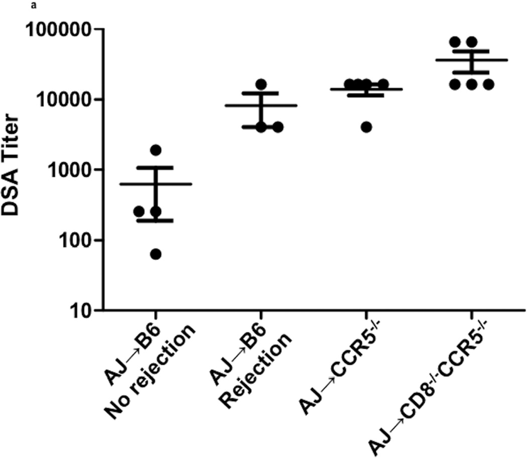
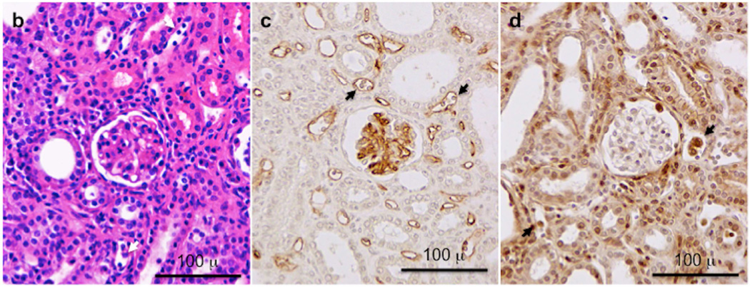
Figure 3. Titers of donor-reactive IgG antibody at the time of kidney allograft rejection in wild type C57BL/6, B6.CCR5−/− and B6.CD8−/−CCR5−/− recipients.
(a) Titers of donor-reactive IgG antibody were determined in serum collected from allograft recipients at the time of graft rejection or from C57BL/7 recipients with long-term surviving allografts collected on day 40 post-transplant. Data indicate individual titers within each recipient group and the mean titer for each recipient group ± SEM. (b–d) Histological evaluation of a rejecting kidney allograft from a wild C57BL/6 recipient on day 36 post-transplant stained with: (b) hematoxylin and eosin; (c) anti-C4d antibody; and (d) anti-Mac2 antibody. Arrows indicate marginating leukocytes. Images shown are representative of 2 individual allografts in the group.


