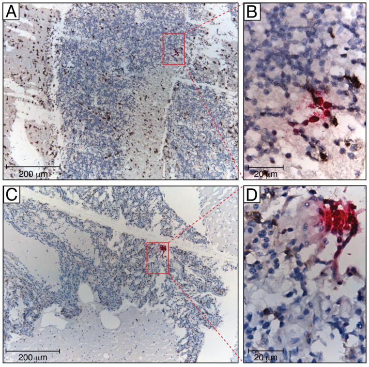
Figure 3. RNAscope of cerebellum in a patient who died with no detectable viral load.
HIV vRNA (coding RNA+, fuchsia) was detected by RNAscope, a novel next generation in situ hybridization technique developed by Advanced Cell Diagnostic, using a HIV gag-pol probe in formalin-fixed and paraffin-embedded cerebellum tissue samples. The RNAscope assay was followed by colorimetric IHC for macrophage markers using mouse mAb to CD163 (Novocastra) and CD68 (Dako) (both brown), and nuclei were counterstained with hematoxylin. To confirm the specificity of in situ hybridization, we used lymph node tissue samples from HIV-negative individual (not shown). Human peptidyl-prolyl cis-trans isomerase B encoded by PPIB gene was detected with the Hs-PPIB probe in the HeLa cell control (ACD) and served as a RNAscope positive control (not shown). Tissue sections were analyzed with a Leica DM6000 B microscope equipped with a Leica DFC 500 camera. Red oblongs in panels A and C outline areas represented in panels B and D, correspondently. Scale bars: 200 μm (A, C) and 20 μm (B, D)

