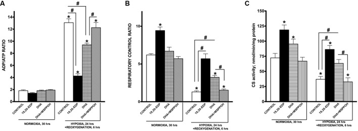
FIGURE 3.
EDPs preserve mitochondrial function following H/R injury. HL-1 cardiac cells were subjected to either 30 h normoxia or 24 h hypoxia and 6 h reoxygenation in the presence of 19,20-EDP (1 μM), DHA (100 μM), and/or MSPPOH (50 μM). Treatment of HL-1 cells with DHA or EDPs during H/R injury sustained (A) the intracellular ratio between ADP and ATP, (B) enhanced and protected mitochondrial respiration and finally, (C) limited the drop in citrate synthase (CS) activity (a marker of mitochondrial content) caused by H/R injury. The ratio between basal and ADP-stimulated respiration is presented as respiratory control ratio (RCR). Activity of CS was used as a marker of mitochondrial content. Values are represented as mean ± SEM; N = 3 independent experiments; ∗p < 0.05 treatment vs. normoxic control, #p < 0.05 treatment group vs. H/R control or DHA/MSPPOH.

