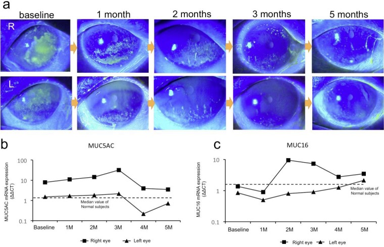
Fig. 2.

Slit-lamp photographs and mucin mRNA expression on the ocular surface of patient 2. a Slit-lamp photographs of the right eye showing the course of corneal fluorescein staining. b Course of MUC5AC mRNA expression on the ocular surface. c Course of MUC16 mRNA expression on the ocular surface. M = Month(s).
