Fig. 1.

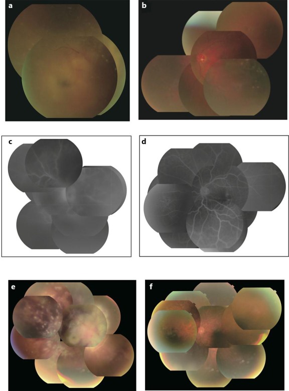
Fundus photographs and fluorescein fundus angiography. a Fundus photographs of the right eye with vitreous opacity, a mild vitreous hemorrhage and extensive multifocal exudation around the optic disc. b Fundus photographs of the left eye with mild vitreous opacity, an optic disc hemorrhage and peripheral white granulomatous exudation. c Fluorescein fundus angiography of the right eye with hyperfluorescence of the optic disc and a wide range of occlusive vasculitis. d Fluorescein fundus angiography of the left eye with retinal vasculitis in the nasal area. e Fundus photographs of the right eye, showing the occlusive vasculopathy with multiple hemorrhages and exudation of the entire retina and retinal fibrosis. f Fundus photographs of the right eye without any sign of apparent progression.
