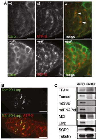
Figure EV3. Larp localization in ovaries and expression of mitochondrial proteins in ovary and somatic tissues.

- Wild‐type and mdi 1 germaria were stained for Larp (green) and ATP‐S (red) to reveal mitochondria. Note that Larp closely associates with mitochondria in wt germarium (arrowheads). Mitochondria in mdi 1 flies completely lack Larp staining. Scale bars, 10 μm.
- Tom20‐LarpGFP (Tom20‐Larp) fusion protein was expressed under the control of nanos‐gal4 in an mdi 1 egg chamber that was stained with ATP‐S (red) to mark mitochondria. Note that Tom20‐Larp is concentrated around mitochondria (ATP‐S) in mdi 1 background. Scale bars, 10 μm.
- Western blots of several mitochondrial proteins in ovary and somatic tissues of wild‐type flies. Boxed are mtDNA replication factors, including TFAM, mtDNA polymerase (Tamas), mitochondrial single‐strand DNA binding protein (mtSSB), mitochondrial RNA polymerase (mtRNAPol), MDI, and Larp. Except for TFAM, most proteins required for mtDNA replication are upregulated in ovary mitochondria. Tubulin was used as a loading control.
