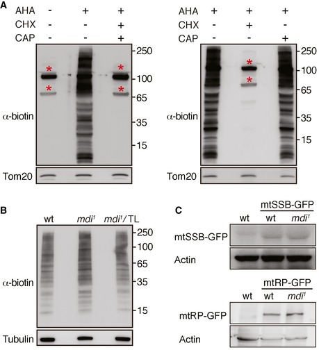
Figure EV5. Western blot analyses of nascent protein synthesis and steady‐state protein levels in ovaries.

- Western blot analyses of nascent protein synthesis in the mitochondrial fraction of the ovary. The nascent protein synthesis was labeled by AHA incorporation and detected by anti‐biotin antibody. Tom20 was used as a loading control. There were two strong bands (*) in the mitochondria fraction without the AHA incubation, indicating two endogenously biotinylated mitochondrial proteins. The AHA signal was mostly blocked in the presence of a cytosolic translation inhibitor, cycloheximide (CHX). Whereas chloramphenicol (CAP) has no impact on the HpG incorporation, indicating the nascent protein synthesis is mainly derived from cytosolic ribosomes associated with mitochondria.
- Western blot analyses of nascent protein synthesis in the cytosolic fraction of wt, mdi 1, and mdi 1 expressing Tom20‐Larp (mdi 1/TL) ovary. Tubulin was used as a loading control. The overall AHA signals indicating the nascent protein synthesis in the three genotypes are comparable.
- Western blot analyses of mtSSB‐GFP and mtRNApol‐GFP (mtRP‐GFP) in wt and mdi 1 ovary. Actin was used as a loading control.
