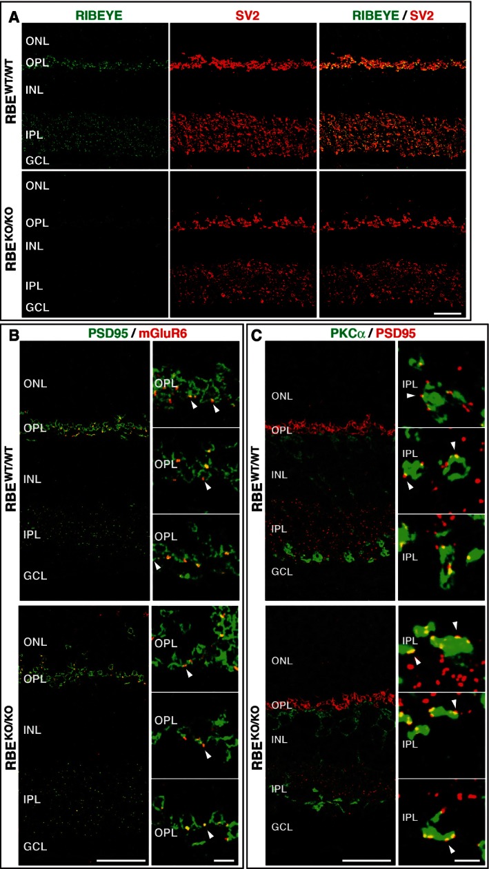
Overview of the synaptic organization of the retina in RBE
WT/WT and RBE
KO/KO mice. Cryostat sections from littermate mice were labeled by double immunofluorescence for the RIBEYE B‐domain (CtBP2; green) and SV2. Scale bar: 20 μm; abbreviations: ONL, outer nuclear layer; OPL, outer plexiform layer; INL, inner nuclear layer; IPL, inner plexiform layer; GCL, ganglion cell layer.
Double immunofluorescence staining of retinas from RBE
WT/WT and RBE
KO/KO mice for PSD95 (green, to label postsynaptic specializations) and mGluR6 (red, to label specifically postsynaptic sites formed by bipolar neurons in photoreceptor synapses). White arrowheads identify mGluR6‐positive puncta at photoreceptor/bipolar cell synapses in the OPL. Abbreviations are as in (A). Scale bars: 20 μm (overview), 5 μm (panel magnification).
Same as (B), except that retinas were stained for PKCα (green, to label bipolar neurons) and PSD95 (red, to label amacrine cells). White arrowheads identify PSD95‐positive puncta at rod bipolar cell synapses in the IPL. Scale bars: 20 μm (overview), 2 μm (panel magnification).

