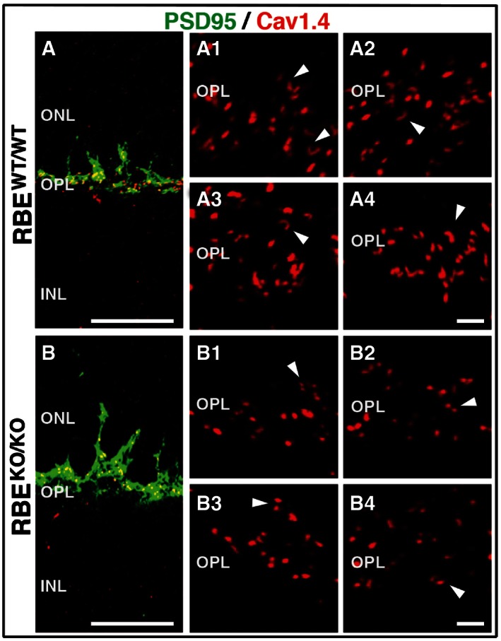
Figure 3. RIBEYE KO results in altered Cav1.4 localization.

-
A, BDouble immunofluorescence staining of retinas from RBE WT/WT and RBE KO/KO mice, respectively, for PSD95 (green, to label presynaptic photoreceptor terminals) and Cav1.4 (red, to label presynaptic Ca2+ channel). Scale bar: 20 μm; abbreviations: ONL, outer nuclear layer; OPL, outer plexiform layer; INL, inner nuclear layer. Magnified images: Magnification of Cav1.4 staining in the outer plexiform layer (OPL) in the retina of (A1–A4) RBE WT/WT and (B1‐B4) RBE KO/KO mice. White arrowheads indicate Ca2+ channel distribution at selected synapses. Images represent 2‐μm z‐stack projections. Scale bar: 2 μm.
