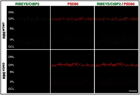
Figure EV3. Immunofluorescence staining of RIBEYE/CtBP2 and PSD95 in wild‐type and RIBEYE KO retina.

Representative double immunofluorescence labeling of RIBEYE/CtBP2 (green, to label the B‐domain common to both splice forms) and PSD95 (red) in semi‐thin sections of RBE WT/WT and RBE KO/KO retina. The RBE KO/KO retina clearly shows a lack of RIBEYE in photoreceptor terminals in the outer plexiform layer and bipolar cell terminals in the inner plexiform layer compared to wild‐type retina. Scale bar: 20 μm; abbreviations: ONL, outer nuclear layer; OPL, outer plexiform layer; INL, inner nuclear layer; IPL, inner plexiform layer; GCL, ganglion cell layer.
