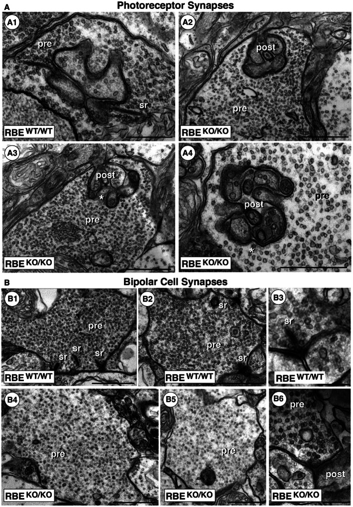
Figure 4. Electron microscopy (EM) shows that RIBEYE KO abolishes synaptic ribbons in retinal synapses formed by photoreceptor and bipolar cells.

- Transmission EM demonstrates the absence of synaptic ribbons in presynaptic terminals of photoreceptor ribbon synapses in RBE KO/KO mice, while synaptic ribbons are readily visible in the terminals of RBE WT/WT mice. Asterisks in (A2) and (A3) denote photoreceptor active zones of RBE KO/KO mice in which the synaptic ribbon is absent. Except for the absence of synaptic ribbons, the ultrastructural appearance of the presynaptic terminals is comparable to that of RBE WT/WT mice. Abbreviations: sr, synaptic ribbon; pre, presynaptic; post, postsynaptic. Scale bars: 500 nm.
- Transmission EM demonstrates the absence of synaptic ribbons in presynaptic terminals of bipolar cell ribbon synapses in RBE KO/KO mice, while synaptic ribbons are readily visible in the terminals of RBE WT/WT mice. Except for the absence of synaptic ribbons, the ultrastructural appearance of the presynaptic terminals is comparable to RBE WT/WT mice. Abbreviations are the same as in (A). Scale bars: 1 μm (B1, B2, B4, B5); 300 nm (B3, B6).
