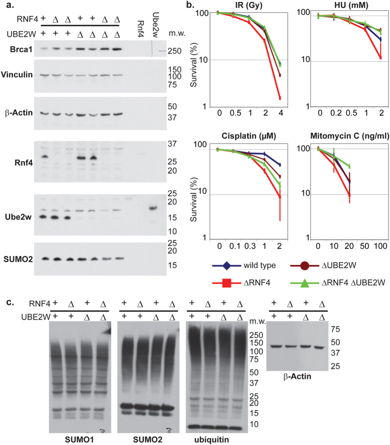
Figure 6. Suppression of ΔRNF4 DNA damage hypersensitivity by ΔUBE2W is conserved in Human.
(a,c) Whole cells extracts of Human HCT116 wild type cells and cells deficient for Rnf4 (ΔRNF4), Ube2w (ΔUBE2W) and Rnf4 and Ube2w (ΔRNF4 ΔUBE2W) were analysed by Western blotting using the indicated antibodies. 3 ng of recombinantly expressed rat Rnf4 and Human Ube2w isoform1 protein were used as a control. Molecular weight marker is indicated on the right inside (kDa). (b) Wild type cells and cells deficient for ΔRNF4; ΔUBE2W and ΔRNF4, ΔUBE2W were subjected to cisplatin, mitomycin C, γ-irradiation and replication stress by HU. The concentration of HU is indicated on the X-axis. Effect on each cell line is indicated of the percentage of colony formation on the Y-axis (logarithmic scale). Data represented as indicated: Wild type (Blue losange), ΔRNF4 (red square), ΔUBE2W (brown circle), ΔRNF4 ΔUBE2W (green triangle). Error bars represent 2 SD.

