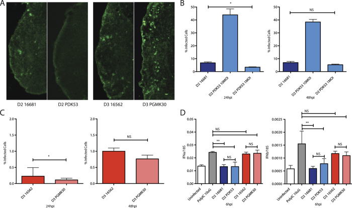
Figure 5. In vivo spread of DENV and adaptation to canine cells.
(A) Immunofluorescence of DENV in popliteal lymph nodes 24 hours after inoculation of virus into the footpads of C57BL6 mice. 1 × 10⁵ pfu of DENV was injected subcutaneously into the footpads of immunocompetent C57BL/6 mice. The draining popliteal lymph nodes were dissected 24 hpi then stained for viral dsRNA (green). (B) Percentage of Madin-Darby Canine Kidney (MDCK) cells infected with DENV-2 as measured by FACS at 24 hpi (left) and 48 hpi (right). At 10MOI, PDK53 infected significantly more MDCK cells (44%) than 10MOI of 16681 (6%). At 1MOI, PDK53 infected fewer cells at 24 hpi (4%), but caught up with 16681 (6%) at 48 hpi. (C) MDCK cells infected DENV-3 at 24 hpi (left) and 48 hpi (right). Both DENV-3 strains infected approximately 1% of cells at both time points. (D) IFNα (left) and IFNβ (right) transcript levels in infected MDCK cells. PDK53, like 16681, did not significantly upregulate interferon expression, unlike infection with either DENV-3 strains or treatment with poly I:C.

