Figure 3.
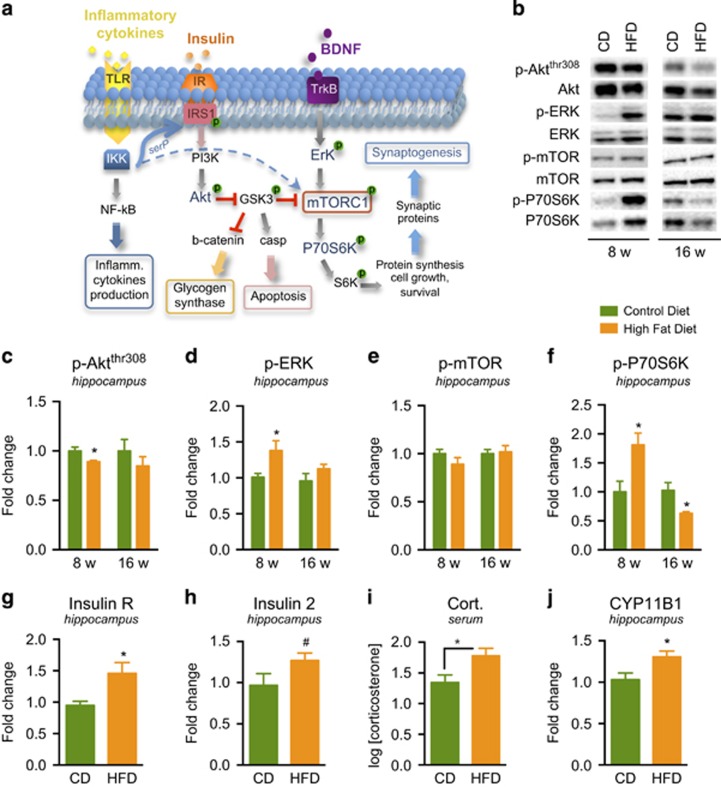
Influence of HFD on insulin, mTORC1 signaling, and corticosterone. (a) Schema showing that activation of insulin receptor (IR) activates Phosphatidylinositol 3 kinase (PI3K) pathway, leading to Akt/mTORC1 signaling pathway involved in protein synthesis required for long-term synaptic plasticity. Phosphorylation (p) of GSK3 at serine 9 leads to the inhibition of GSK3 activity. Levels of the active form of GSK3 (dephosphorylated) are increased in depression, diabetes, and Alzheimer's disease. (b) Representative immunoblots obtained after 8 and 16 weeks of CD or HFD. (c–f) Quantitative results of phospho (p) protein immunoblots are based on total levels of protein for each particular kinase. HFD differentially alters the phosphorylation of Akt, ERK, and P70S6K in the hippocampus, determined by western blot analysis but no change was detected in levels of mTOR. (g and h) Levels of insulin receptor and insulin 2 mRNA in hippocampus were determined by qPCR analysis: HFD (16 weeks) significantly increased insulin receptor mRNA and tended to increase insulin 2 mRNA. (i) HFD for 16 weeks also increased serum corticosterone levels (log-transformed data), and (j) hippocampal levels of CYP11B1 mRNA. Results are presented as the mean±SEM, n=8 per group. *P<0.05 compared with the control diet group (two-tailed Student's t-tests), #P=0.08. CYP11B1: cytochrome P450, family 11, subfamily B, polypeptide 1.

