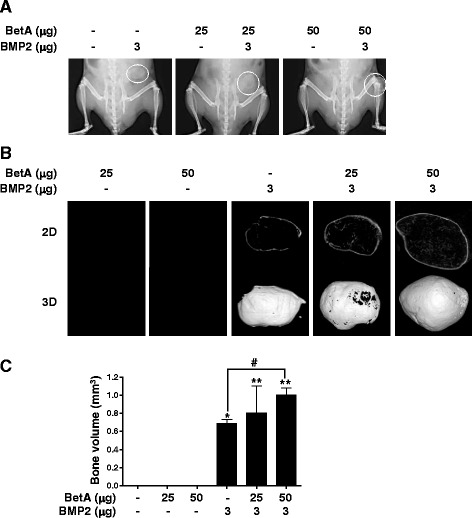
Fig. 4.

Radiographic study of the ectopic bone formation by BetA, BMP2 and BetA/BMP2 composite implants. BetA (25 or 50 μg) with or without BMP2 (3 μg) were administered with absorbable collagen sponges into the subcutaneous spaces in the back of mice. After 4 weeks, microradiographic analyses were performed. a Soft X-ray analysis. Ectopic bone formation was seen in BMP2- or BetA/BMP2-treated groups. Dotted circles in the right side of mice indicate the new ectopic bones. b Micro-computed tomography analysis. Each ectopic bone of (a) was isolated, and then scanned by μ-CT. The image slices were reconstructed three dimensionally as in Materials and Methods section. c Quantitative analysis. Ectopic bone volume was measured using a CT-Analyzer program. *, p < 0.05, and **, p < 0.01 compared to the control group (collagen sponge alone). #, p < 0.05 compared to the indicated group. Representative data are shown. n = 5
