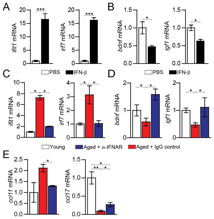
Fig. 3. Restored CP function after neutralization of the age-induced type I IFN response in the brain.

(A–B) ifit1, irf7 (A), and bdnf and igf1 (B) mRNA expression in CP epithelial cells cultured for 24h with murine IFN-β (n=4 per group; bars represent mean ± SEM; *, P < 0.05; ***, P < 0.001, Student’s t test). (C) ifit1 and irf7 mRNA expression in the CP 3 days after α-IFNAR or IgG control i.c.v. administration. (D–E) bdnf and igf1 (D) and ccl11 and ccl17 (E) mRNA expression in the CP 7 days after α-IFNAR or IgG control i.c.v. administration (n=6–7 per group). Throughout the figure, bars represent mean ± SEM; *, P < 0.05; **, P < 0.01; one-way ANOVA with Newmann-Kleus post-hoc test.
