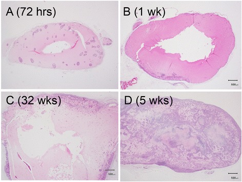
Fig. 2.

Morphology of MCF-7 xenografts at time points post implantation. The paraffin-embedded, sectioned tissue slices with hematoxylin & eosin staining to visualize morphology were examined by bright field microscopy. a Tumor masses were grown as enlarged cysts with numerous adenoid rosette structures with tumor cell groups distributed inside the matrigel wall at 72 h post implantation. b, c The tumor cells gradually proliferated and aggregated near the exterior edge of the matrigel wall at 1–3 weeks post implantation. d The xenografts developed as solid masses with central necrosis after 5 weeks post implantation
