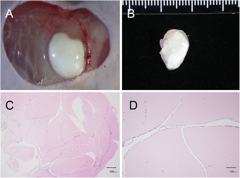
Fig. 7.

Morphology of the developed (5-week) benign xenograft. There were no noticeable newly-formed blood vessels on the surface of either the xenograft in situ (a) or in the harvested tissue (b) or inside the developed mass with histological H&E staining (c). The benign masses appeared as pink-colored blocks and were infiltrated by fibrotic cells exhibiting cleft- or slit-like openings inside the xenografts (c, d)
