
Fig. 3.
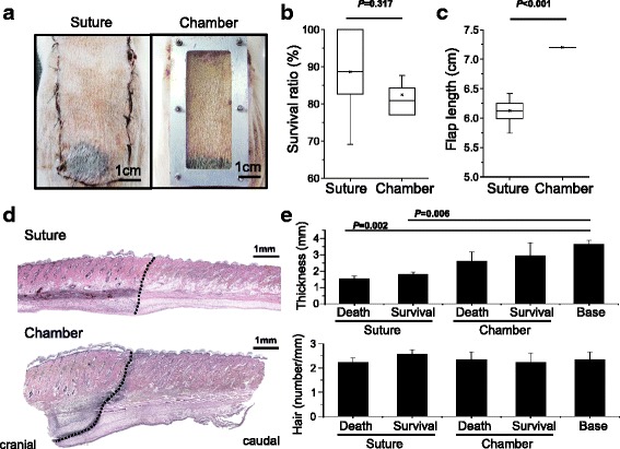
Reproducible skin survival by flap chamber. Gross picture of flap survival between suture and chamber model (a). The box plot of survival ratio distribution of dorsal flap (n = 8 in each group) (b). The skin contraction decreased the flap length in the suture model but not in the flap chamber (c). Hematoxylin-and-eosin staining from cranial to caudal, across the skin necrosis line (black dotted line) (d). Quantification of skin thickness and number of hair follicles were compared from cranial to caudal survival and death region (e)