Figure 3.

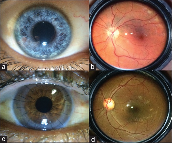
Examples of normal (a) anterior and (b) posterior segment images and of subtle findings including (c) detail of all sutures in corneal transplant and (d) optic nerve cupping

Examples of normal (a) anterior and (b) posterior segment images and of subtle findings including (c) detail of all sutures in corneal transplant and (d) optic nerve cupping