Figure 8.

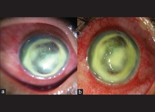
Image of corneal abscess taken with (a) anterior chamber EyeGo imaging and (b) slit lamp imaging shows similar level of detail

Image of corneal abscess taken with (a) anterior chamber EyeGo imaging and (b) slit lamp imaging shows similar level of detail