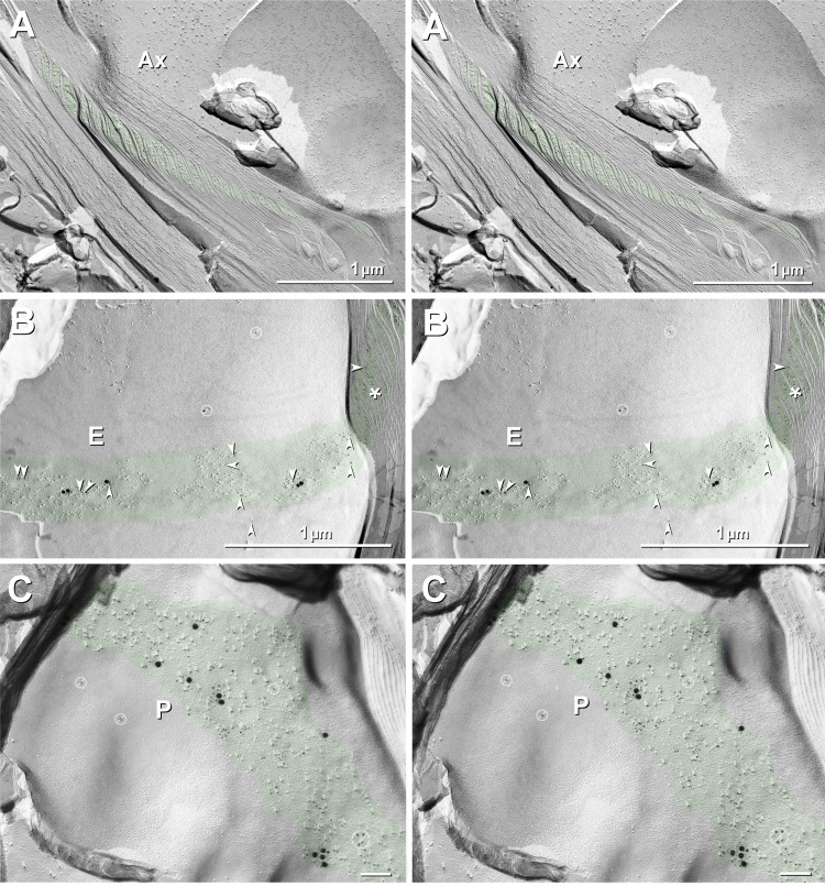
Fig. 11.
Cross fracture (A) and en face fractures (B and C) through SLIs (green overlays) from dual-double-labeled sciatic nerve of Cx32 ko mouse. A: cross-fractured SLIs are only weakly immunogold labeled for Cx29. B and C: fractures that include en face views of one of the steps in the SLI E-face (B) and P-face (C) are strongly labeled for Cx29 [5-nm (arrowheads) and 20-nm gold beads], but they are not at all labeled for KV1 (10- and 30-nm gold beads; none present in either E- or P-face views). (See Figs. 2 and 4 for immunofluorescence imaging correlates.) E-face labeling represents “cryptic” labeling of Cx29 in the subjacent membrane of another step in the SLIs. No rosettes are present in E- or P-faces of SLIs in either formaldehyde-fixed (present study) or glutaraldehyde-fixed tissue (see also Stolinski et al. 1985), suggesting that Cx29 is present as singlet IMPs that do not form rosettes without KV1.1-containing coupling partners. Gold beads in barred circles in B and C are identified as “noise” because they are on top of the replica, where no labeling is possible. Green overlays (and asterisk in B) mark cytoplasmic expansions that characterize the SLIs. Scale bar, 0.1 μm (C).

