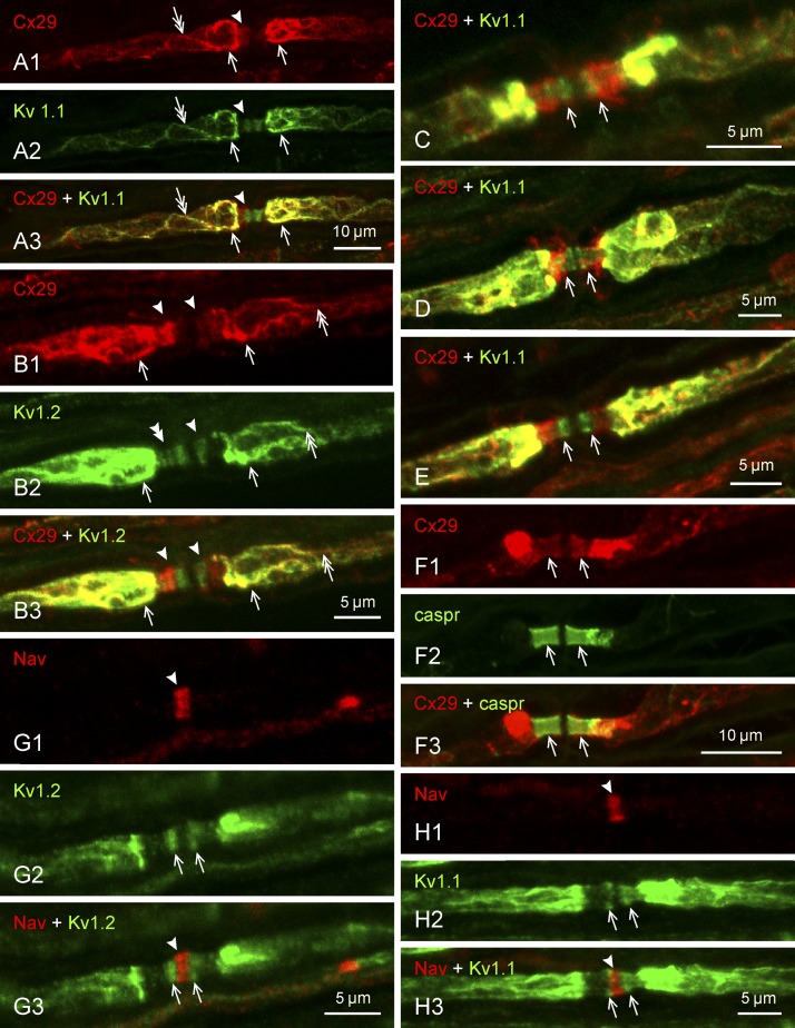
Fig. 3.
Immunofluorescence labeling of Cx29 and KV1.1 at regions surrounding nodes of Ranvier in adult rat sciatic nerve. Proteins labeled and color code for labeling are indicated at top left of each panel. Images in panels with the same lettering show the same field. A: labeling for both Cx29 (A1) and KV1.1 (A2) is dense in the juxtaparanodal region (arrows), faint in the paranodal region (arrowheads), and can be seen extending up to and into the juxtaparanodal region along the inner mesaxon (double arrows), with overlap of labeling in most of these regions (A3). B: similar colocalized labeling of Cx29 (B1 and B3) in relation to KV1.2 (B2 and B3) is shown at juxtaparanodal (arrows), paranodal (arrowhead), and mesaxonal (double arrows) regions. At paranodes, labeling of KV1.2 is resolved as several transverse bands (B2, double arrowhead). C–E: overlays of labeling for Cx29 and KV1.1 at the paranodal/juxtaparanodal region, showing variable degrees of overlap between the transverse bands of KV1.1 labeling at paranodes and dense (C), moderate (D), or weak (E) labeling for Cx29 (arrows) extending into this region. F: labeling of Cx29 in the paranodal area (F1, arrows) overlaps with labeling for the paranodal marker caspr (F2, arrows), as shown in overlay (F3, arrows). G and H: labeling for KV1.2 (G1) and KV1.1 (H1) at paranodes (arrows) does not extend into nodes of Ranvier, identified by their labeling of sodium channels (NaV) (G2 and H2, arrowheads) sandwiched between the paranodal transverse KV bands (G3 and H3). Scale bars are as indicated on each lettered group of panels.

