Fig. 4.
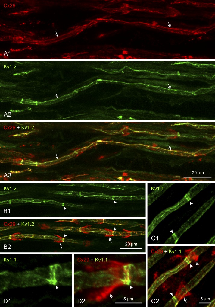
Immunofluorescence double labeling of Cx29 and KV channels along myelinated axons in sciatic nerve of adult rat. Proteins labeled and color code for labeling are indicated at top left of each panel. Images in panels with the same lettering show the same field. A: magnification of internodal regions, showing both Cx29 (A1) and KV1.2 (A2) localized at myelinated fibers as a single near-continuous linear strand of labeling (arrows) running along the internodal segment in an area overlying the axon, with Cx29/KV1.2 colocalization along these strands shown in overlay (A3, arrows). B: internodal regions of myelinated fibers, showing KV1.2 localized as intermittent narrow transverse bands along axons (B1, arrowheads) and Cx29 at funnel-shaped SLIs (B2, arrows), with the KV1.2 bands situated at the narrow ends of the Cx29-immunopositive incisures (B2, arrowheads). C and D: magnifications of SLIs, showing labeling for KV1.1 localized as transverse bands along axons (C1, arrowheads), with the bands consisting of a doublet of bars (arrowheads) overlapping with the narrow end of the incisures labeled for Cx29 (C2, arrows), as shown in overlay (arrowheads) and at higher magnification (D). Scale bars are as indicated on each group of panels.

