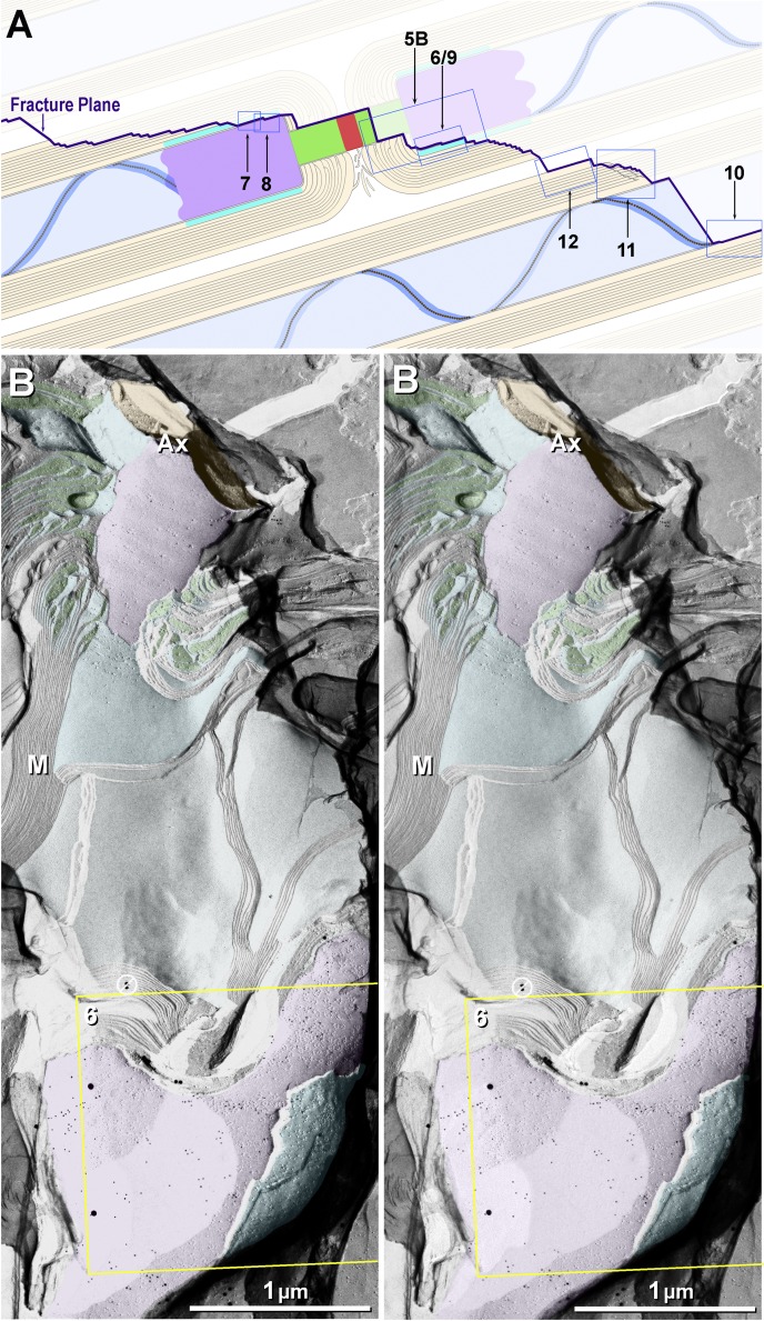
Fig. 5.
Eight of the 10 principal fracture planes examined in this study (A) and stereoscopic low-magnification image from dual-double-labeled freeze-fracture replica immunogold labeling (FRIL) replica from mouse sciatic nerve revealing the paranodal and juxtaparanodal membranes (B). A: 2 freeze-fractured myelinated axons (blue axoplasm), with the fracture plane (purple line) exposing a node of Ranvier, E-face of the paranodal axolemma, E-face of the juxtaparanodal axolemma (JPAX), and P-face of juxtaparanodal innermost myelin (JPIM; box 6/9), pitted E-face of juxtaparanodal myelin (box 7), P-face of the JPAX (box 8), E- and P-faces of inner mesaxonal myelin (IMAX; blue overlay and box 10), Schmidt-Lanterman incisures, and P-face of outer surface of myelin. Boxes correspond to numbered figures. For simplicity, only 10 of the usual 25–50 layers of myelin are indicated. Dark aqua and box 8, KV1.1-enriched JPAX; blue ribbon and box 10, IMAX; light blue, axon; beige, myelin; red, node; green, paranodes; purple, juxtaparanodes. B: low-magnification overview of the paranodal and juxtaparanodal membranes in a single axon; boxed area is shown at higher magnification in succeeding figures. At this magnification, 3 of the 4 sizes of gold beads used for dual double labeling are discernible (10- and 30-nm gold for KV1.1 and 20-nm gold for Cx29) inside the yellow box. Abundant 5-nm gold beads (also for Cx29) cannot be discerned but are detectable when box 6 is further enlarged as Fig. 6. A pair of gold beads (barred circles at top edge of yellow box) on top of the replica are positively identified as “noise.” Purple overlay at top is axolemmal E-face, with impressions of paranodal loops of myelin; purple overlay at bottom is E-face of the JPAX region within the same complexly fractured myelin sheath (M). Aqua overlays (middle and bottom) are portions of the underlying P-face of innermost myelin, including tips of paranodal loops. Green overlays are cytoplasm of paranodal loops. Local area “dodging” was used to reduce the photographic intensity of superimposed replica fragments and areas of folded replica. Ax, axoplasm. Scale bar, 1 μm.

