Fig. 6.
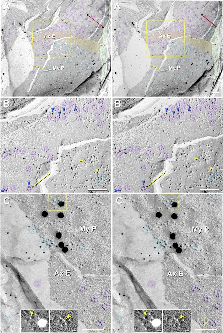
Stereoscopic images of particle rosettes in JPAX E-face and JPIM P-face from Cx32 ko mouse (A and B) and from WT mouse (C). A and B: E-face rosettes (purple overlays) in the axolemma are labeled for KV1.1, and P-face rosettes (aqua overlays) in innermost myelin are labeled for Cx29. Four sizes of gold beads are used: 2 for KV1.1 (10- and 30-nm gold) and 2 for Cx29 (5- and 20-nm gold). Double-ended red arrow compares 30- vs. 20-nm gold beads; double-ended yellow arrow compares 10- vs. 5-nm gold beads. Green overlays are tongues of myeloplasm at inner mesaxon. Apposing strips of axolemmal E-face (AxE; orange overlay) and myelin P-face (MyP; yellow overlay) are deficient in rosettes, whereas rosettes are abundant on either side of this band. B: High-magnification stereoscopic image of AxE (enlarged from yellow box in A). Most axonal rosettes are labeled by one to four 10-nm gold beads (blue arrowheads) for KV1.1. In bottom right quadrant, the fracture plane dropped from the AxE to the MyP, where the rosettes and clusters of 9-nm particles are labeled for Cx29 by 5-nm gold beads (yellow arrowheads). Purple overlays are rosettes of axolemmal 9-nm E-face particles. Rosettes of P-face particles (aqua overlays) in innermost myelin are labeled for Cx29. C: fracture from AxE to MyP in a WT sample that was dual-double-labeled for Cx29 (∼18 10-nm and six 30-nm gold beads) and for cytoplasmic epitopes of KV1.1 (5- and 20-nm gold; none present). As shown here, cytoplasmic epitopes of KV1.1-containing particle rosettes were never labeled. Purple overlays are unlabeled axonal rosettes. Aqua overlays are clusters and rosettes of 9-nm intramembrane particles (IMPs) labeled for Cx29. Left inset, myelin P-face particles labeled for Cx29 (from top yellow box), presented with black shadows to reveal the central “dimples” (yellow arrowhead). Right inset, axolemmal E-face rosette (from bottom yellow box), also presented with black shadows to reveal the central dimple in each KV1.1-containing IMP (yellow arrowhead). Scale bar, 0.1 μm (10 nm in insets).

