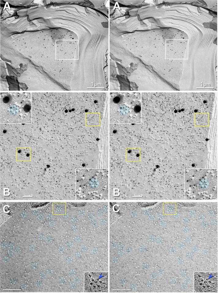
Fig. 7.
Stereoscopic images of labeled JPIM P-face (A and B) and unlabeled JPIM E-face (C). A: high labeling specificity, high labeling efficiency (LE; >1:3), and high signal-to-noise ratio for Cx29 in innermost adaxonal myelemma of a large-diameter axon in mouse sciatic nerve. Box in A is further magnified as B. Slight local area dodging was used to reduce the photographic intensity of the clump of antibodies outside the top left region of the box. B: at higher magnification, the excessively high LE is shown to result in potential obscuring of some particles. Yellow boxes are enlarged (insets) to reveal the greater LE for small gold beads. C: E-face imprints of densely packed Cx29 rosettes (aqua overlays) in JPIM of glutaraldehyde-fixed (i.e., not labeled) sciatic nerve from WT mouse. Most rosettes are complete rings, but a few incomplete rings contain only 4 or 5 pits. Most pits contain a central peg (blue arrowhead in inset; shown with black shadows). Pegs represent the frozen water-filled matrix extracted from the ion channel during cleaving (Hirokawa and Heuser 1982; Rash et al. 2004a), implying that most Cx29 channels are open when fixed. Paucity of E-face IMPs indicates the virtual absence of all other types of ion channels in E-faces. Likewise, in the complementary P-faces (B), there are few, if any, particles other than Cx29. Scale bars, 1 μm (A), 0.1 μm (B and C), and 10 nm (B and C, insets).

